- HOME
- Research
Research
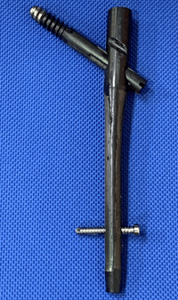
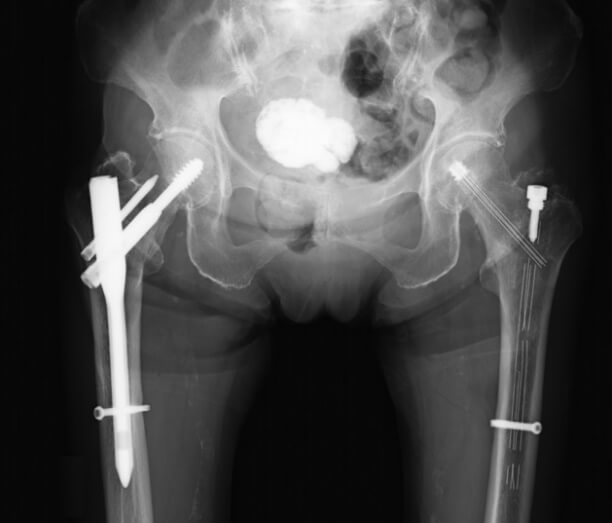
1.Development of devices for osteosynthesis and artificial joints using carbon-fiber-reinforced polyetheretherketone (CFR/PEEK)
Carbon-fiber-reinforced polyetheretherketone (CFR/PEEK) is a new material that has high fatigue strength and low rigidity. We have developed the devices for osteosynthesis based on CFR/PEEK for the treatment of proximal femoral fracture. We have also investigated a new technique for biocompatibility of CFR/PEEK and developed new-generation artificial joints based on CFR/PEEK.
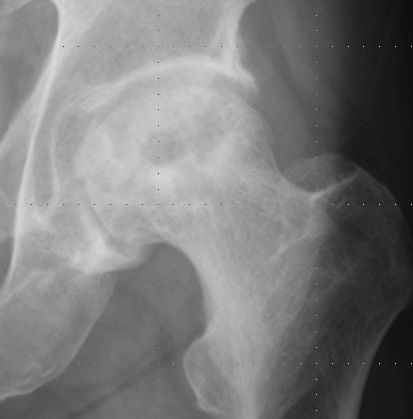
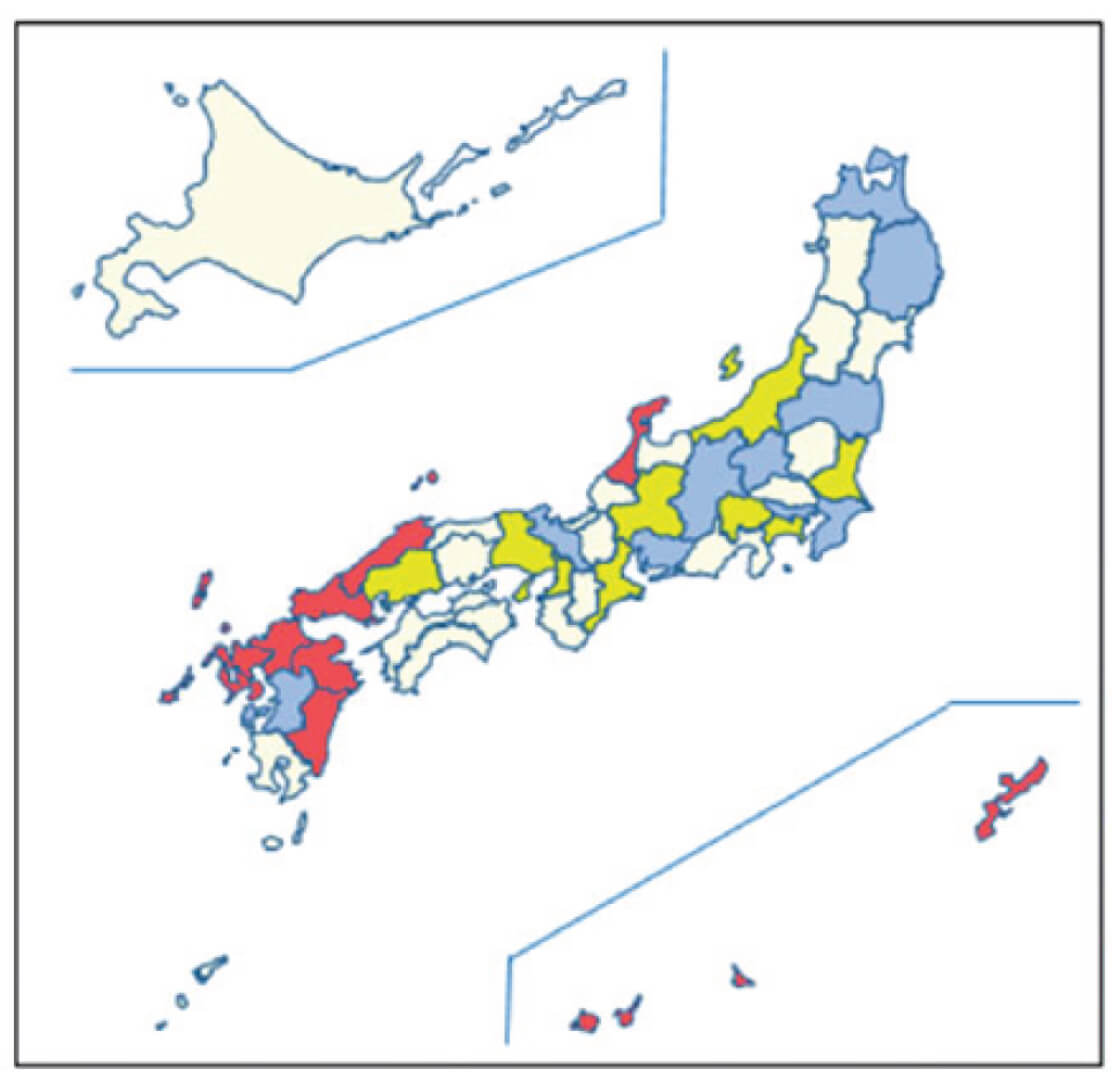
2.Epidemiology and clinical guidelines for osteonecrosis of the femoral head (ONFH)
ONFH is one of the causes of acute hip pain in the early stage and often leads to the progressive collapse of the femoral head with joint destruction. The pathomechanism and cause of ONFH has not been clarified, and ONFH has been recognized as a designated intractable disease in Japan. Prof. Sugano is the president of the Japanese Investigation Committee (JIC) for ONFH under the auspices of the Ministry of Health, Labor, and Welfare and has led the epidemiological study for ONFH with JIC members. The JIC members also developed the clinical guidelines for ONFH in 2019.
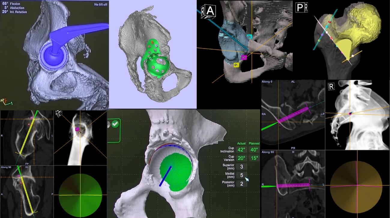
3.Clinical Application of Navigation and Robotics to Hip Surgery.
We have been using the CT-based navigation system to perform all hip surgeries, including primary total hip arthroplasty, revision hip arthroplasty, periacetabular osteotomy, proximal femoral osteotomy, hip arthroscopy, and pelvic fracture treatment. The accuracy and its determining factor are evaluated to improve its performance and user-friendliness. In 2018, we introduced the robot-arm assisted system for total hip arthroplasty (MAKO Total Hip, Stryker) first in Japan. We have started a multi-center study to evaluate its accuracy and usefulness in total hip arthroplasty.
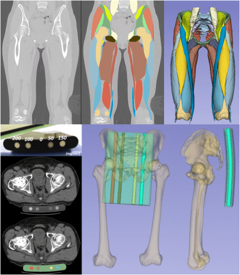
4.Application of artificial intelligence to the hip.
With a collaboration with the Nara Institute of Science and Technology (Prof. Sato, Assoc. Prof. Otake), we have been applying artificial intelligence (AI) to segment the bones, muscles, and calibration phantom from clinical CT images. Further, we are now applying AI to classify the CT images that are gathered as a big data project at the National Institute of Informatics.
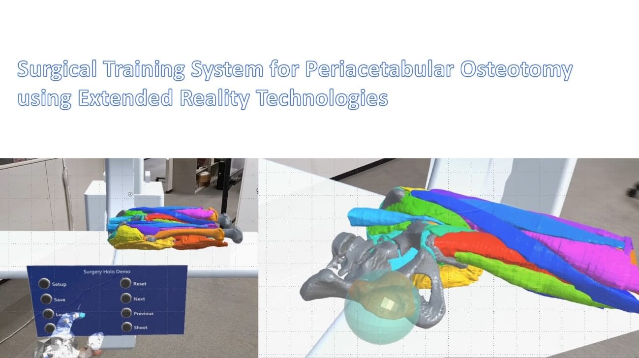
5.Development of the Novel Surgeon Training System using Extended Reality Technology
We are developing the novel surgical training system using extended reality technology. It is a more realistic system that includes various levels of surgical difficulty and anatomical variations. This is a collaborative study with Prof. Hirokazu Kato of Nara Institute of Science and Technology
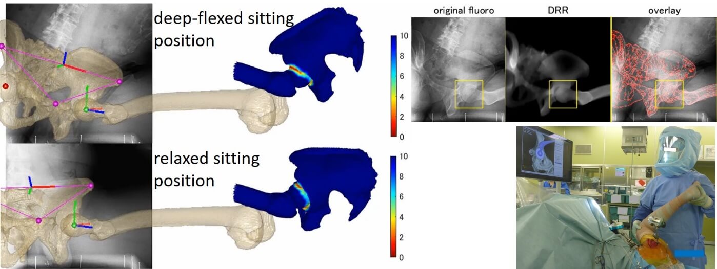
6.Application of novel technology to the analysis of spinopelvic biomechanics
We are developing an application of the motion analysis that combines individual skeletal models with motion capture or X-ray fluoroscopic video to analyze spinopelvic biomechanics. We are also developing a clinical application of motion analysis using wearable sensors and multi-video data to surgical planning or perioperative evaluation.
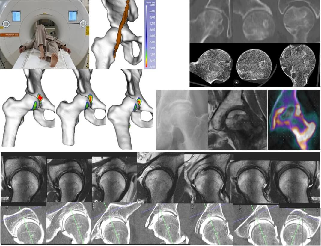
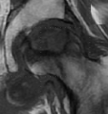
7.Application of novel imaging technology to the analysis of the pathogenesis of hip disease
We are conducting further analyses for the pathology of hip diseases using large-diameter MRI for the dynamic analysis of the iliopsoas muscle, arthrography CT and MR radial imaging for the evaluation of hip joint cartilage and labrum, micro-CT evaluation for the microstructure of the femoral head, and SPECT-CT for the three-dimensional evaluation of blood flow in the femoral head.